



DNA Stain
There are several different stains that can be used to visualize and photograph DNA after separation by gel electrophoresis. Molecules of the dye adhere to DNA strands and fluoresce under UV light, showing where the bands are within the gel.
Hoechst stains are part of a family of blue fluorescent dyes used to stain DNA.There are three related Hoechst stains: Hoechst 33258, Hoechst 33342, and Hoechst 34580. The dyes Hoechst 33258 and Hoechst 33342 are the ones most commonly used and they have similar excitation/emission spectra.
All Product Categories
-
DNA Stain Related Products (91)
Related Products (91)

-
MeCY5-NHS ester triethylamine
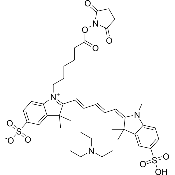 0 ImagesCat. No.: HY-135413BSynonyms: Sulfo-Cyanine5 NHS ester triethylamineMeCY5-NHS ester (potassium) is a reactive dye and can be used for labeling protein nucleic acid.
-
Azure A chloride
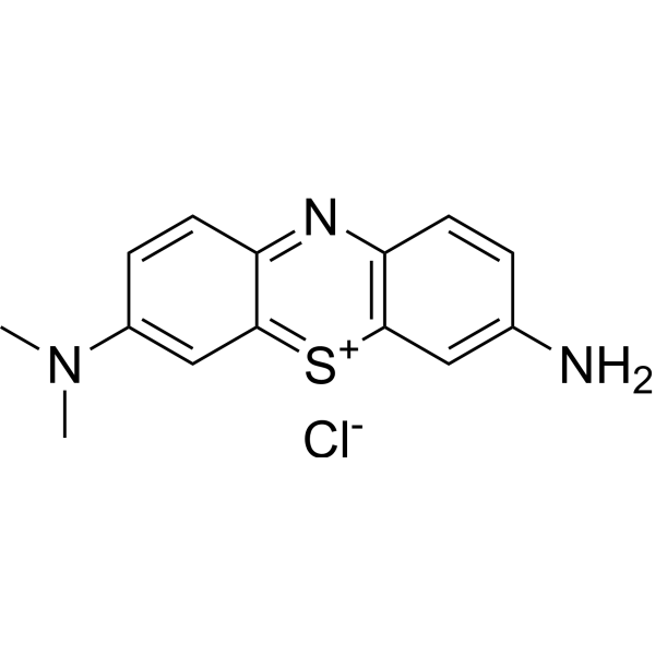 0 ImagesAzure A chloride is a phenothiazine dye. Azure A chloride is an alternative DNA dye used for the separation of DNA and protein fragments in agarose gel electrophoresis and PAGE. Azure A chloride can be chemisorbed on the surface of mild steel according to the Langmuir adsorption isotherm to form a protective film. Azure A chloride binds to double-stranded DNA in a non-cooperative manner via weak intercalation, triggering molecular conformational disturbance, restricted rotational motion, and changes in optical activity.
-
Methyl Green
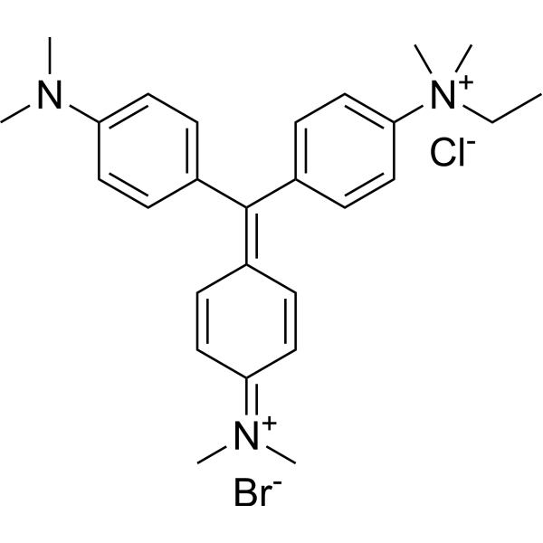 0 ImagesCat. No.: HY-D0163CAS No.: 14855-76-6Methyl Green is a non-intercalating fluorescent labeling agent that selectively binds to the major groove of DNA. Methyl Green electrostatically interacts with the major groove of DNA through positively charged groups, exhibiting key activities such as high affinity, resistance to photobleaching, and stable fluorescence emission. Methyl Green can be directly measured by microscopy and flow cytometry, with peaks at 633 and 677 nm. Methyl Green can be used for fluorescent labeling of the nuclei of embryonic tissues or cells, or DNA staining and cell activity detection in gel electrophoresis[1][2][3].
-
G-quadruplex DNA fluorescence probe 1
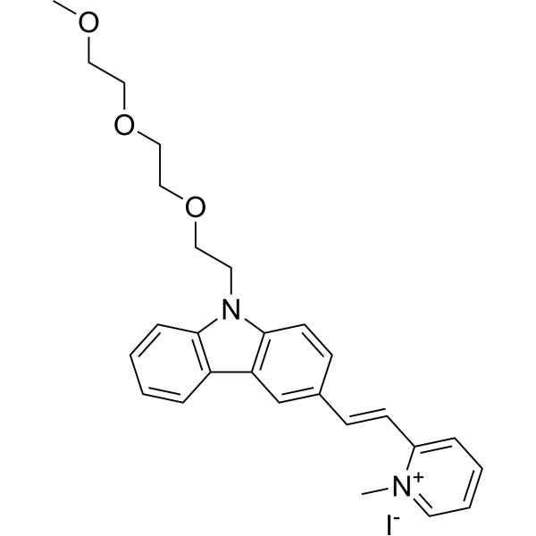 0 ImagesCat. No.: HY-147652G-quadruplex DNA fluorescence probe 1 (Compound E1) is a selective G-quadruplex DNA targeting fluorescent probe. G-quadruplex DNA fluorescence probe 1 can pass through membrane and enter living cells with low cytotoxicity.
-
Cyanine 3 Tyramide
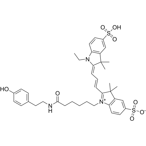 0 ImagesSynonyms: Tyramide-Cy3Cyanine 3 Tyramide (Tyramide-Cy3) is an orange fluorescent dye used as a reporter fluorescent substrate for horseradish peroxidase (HRP)-catalyzed deposition, which serves as a signal amplification technique in immunoassays and in situ nucleic acid hybridization.
-
Cyanine 5 Tyramide
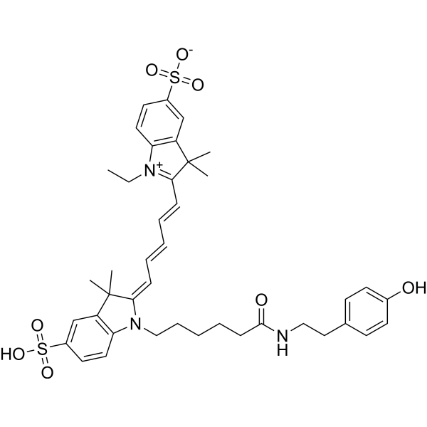 0 ImagesCat. No.: HY-136247CAS No.: 1431148-26-3Synonyms: Tyramide-Cy5Cyanine 5 Tyramide (Tyramide-Cy5), a red fluorescent dye, is utilized as reporter fluorescent substrate for horseradish peroxidase (HRP)-catalyzed deposition that is signal amplification technique in immunoassay and in situ hybridization of nucleic acids.
-
Hoechst 33258 analog
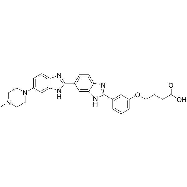 0 ImagesHoechst 33258 analog is a marker dye in Hoechst series. Hoechst is A live nuclear marker dye. Hoechst binds to the grooves in the DNA double strand, which tends to be A/ T-rich DNA strand. Although it binds to all nucleic acids, the A/ T-rich double strand DNA significantly enhances fluorescence intensity Therefore,Hoechst dye can be used for living cell labeling. The fluorescence intensity of Hoechst dye increases with the increase of pH of solution.
-
LDS-751
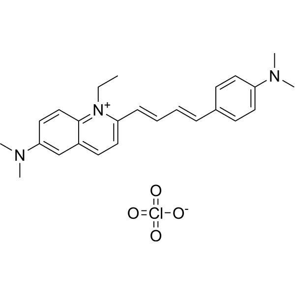 0 ImagesLds-751 is a nucleic acid stain that mainly detects DNA. Lds-751 is a nucleic acid stain that mainly detects DNA. Lds-751 has a high affinity for DNA and fluorescence is enhanced after binding, but the maximum emission wavelength is 670nm. Lds-751 and Thiazole orange can be used for the differentiation of red blood cells, platelets, reticulocytes, and nucleated cells and can be stimulated at 488nm. Studies have shown that LDS-751 binds almost exclusively to mitochondria when incubated with nucleated living cells. After nucleated Acridine Orange (HY-101879) staining and LDS-751 treatment of cells, confocal microscopy revealed almost no co-location of the cells. Staining with Rhodamine 123 (HY-D0816), a dye known to bind polarized mitochondria, was almost identical to the pattern observed with LDS-751.
-
Ethidium homodimer
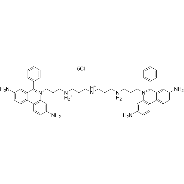
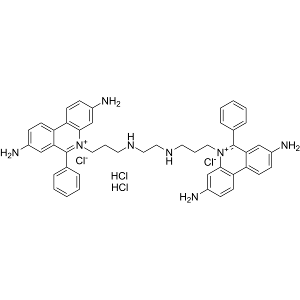 0 ImagesSynonyms: EthD-1Ethidium homodimer (EthD-1) is a high-affinity fluorescent nucleic acid dye commonly used to stain mammals, bacteria, yeast, and fungi. Ethidium homodimer binds to DNA or RNA, enhancing fluorescence more than 30 times. The Ethidium homodimer has a strong positive charge, so it cannot cross cell membranes and stain living cells; But the Ethidium homodimer can cross the disordered region of the dead cell membrane to reach the nucleus and embed the DNA double strand to produce red fluorescence. Therefore, Ethidium homodimer is a relatively sensitive nucleic acid stain that can accurately detect nucleic acids in solution or in decomposing cells. Ethidium homodimer binds DNA, Ex/Em=528/617 nm.
-
NIR-Red Dead Cell-1 Dye
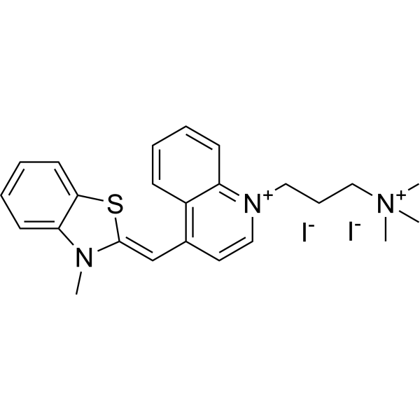 0 ImagesCat. No.: HY-D0917CAS No.: 157199-59-2NIR-Red Dead Cell-1 Dye is a DNA-binding fluorescent dye for non-living cells (Ex/Em=515 nm/531 nm). NIR-Red Dead Cell-1 Dye can intercalate into base pairs of double-stranded DNA and produce stronger fluorescence. NIR-Red Dead Cell-1 Dye is suitable for necrotic cells or late apoptotic cells with damaged cell membranes, showing green fluorescence under fluorescence microscopy or flow cytometry. NIR-Red Dead Cell-1 Dye can be used to distinguish live cells from dead cells and distinguish cell membrane integrity. NIR-Red Dead Cell-1 Dye can be attached to the surface of Feraheme (FH) nanoparticles (NPs) to obtain fluorescent dye-functionalized NPs for drug delivery studies.
-
1-Nitropyrene
 0 ImagesSynonyms: 3-Nitropyrene1-Nitropyrene is a DNA probe whose metabolites can form nucleic acid adducts with DNA.
-
Ethidium bromide (solution)
 0 ImagesEthidium bromide (solution) is an intercalating agent commonly used as a fluorescent tag (nucleic acid stain) in molecular biology laboratories for techniques such as agarose gel electrophoresis.
Solvent and Concentration: Sterile water: 10 mg/Ml
-
Biotin-16-dUTP trisodium
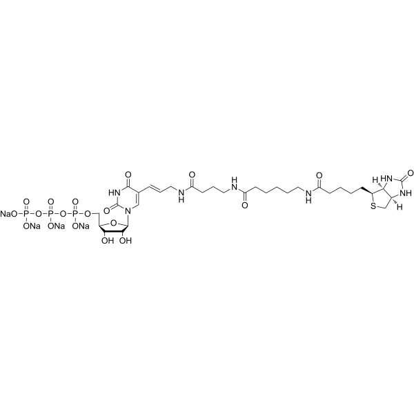
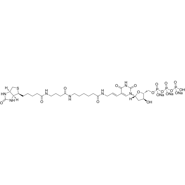 0 ImagesCat. No.: HY-D1022APurity: 99%Synonyms: Biotin-16-deoxyuridine-5'-triphosphate trisodiumBiotin-16- dUTP (Biotin-16-deoxyuridine-5'-triphosphate) trisodium can be used to replace its natural counterpart dTTP by enzymatically incorporating it into DNA/cDNA. Biotin-16- dUTP trisodium can be used to produce biotinylated DNA probes in a variety of assay applications.
-
6-TAMRA free acid
 0 ImagesCat. No.: HY-W142395CAS No.: 150322-06-86-TAMRA free acid is a fluorophore widely used in the preparation of fluorescent antibodies and avidin derivatives for immunohistochemistry.
-
JOE
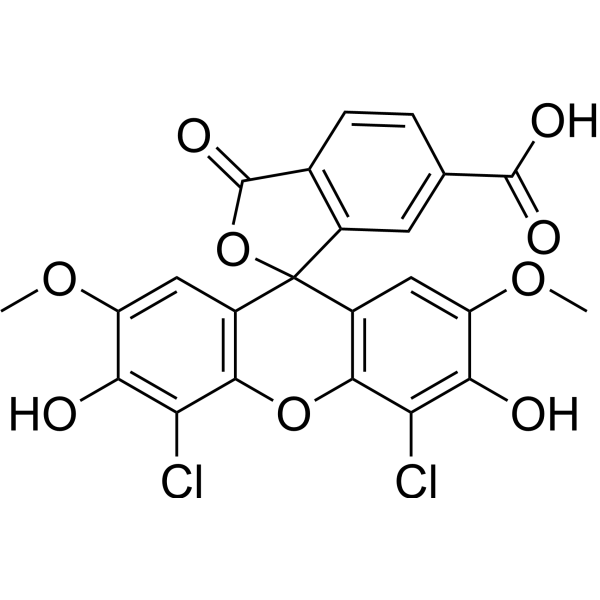 0 ImagesCat. No.: HY-D1090CAS No.: 82855-40-1JOE is a xanthene fluorophore (i.e., 4′,5′-dichloro-2′,7′-dimethoxy-5 (6)-carboxyfluorescein; 2',7'-dimethoxy-4',5'-dichloro-6-carboxyfluorescein) with an absorption wavelength of approximately 525 nm and an emission wavelength of approximately 550 nm. The fluorescence quantum yield of JOE correlates with the rigidity of the linker arm and the distance to dG nucleoside. JOE is commonly used as a fluorescent label for oligonucleotides and molecular beacon probes, and also serves as the acceptor fluorophore in fluorescence energy transfer primers for DNA sequencing.
-
DFHBI-1T (solution)
 0 ImagesCat. No.: HY-DY1047CAS No.: 1539318-36-9DFHBI-1T (solution) is a membrane-permeable RNA aptamers-activated fluorescence probe (ex/em=472 nm/507 nm). DFHBI-1T binds to RNA aptamers (Spinach, Spinach2, iSpinach, and Broccoli) and causes specific fluorescence and lower background fluorescence. DFHBI-1T is used to image RNA in live cells.
Solvent and concentration: DMSO: 20 mM
-
5(6)-ROX
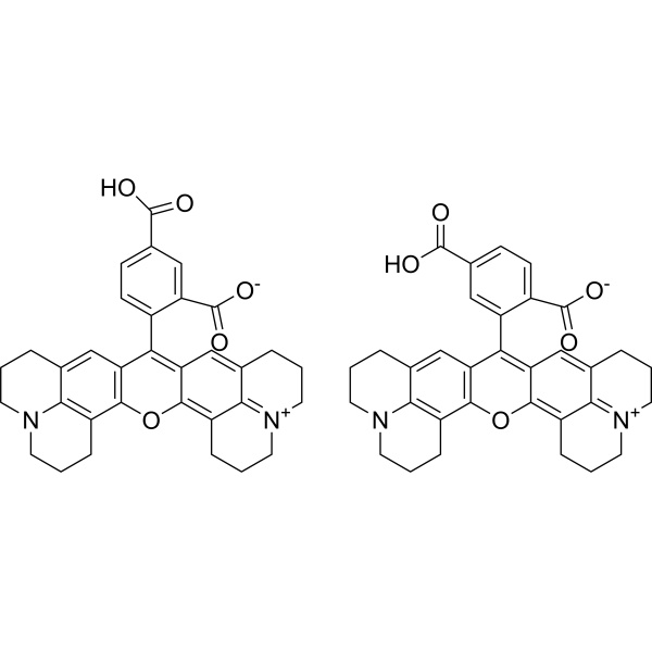 0 ImagesSynonyms: 5(6)-Carboxy-X-rhodamine5(6)-ROX (5(6)-Carboxy-X-rhodamine) is a nucleic acid fluorescent label which can be used as a reference dye for real-time polymerase chain reaction (Em/Ex = 605/585 nm).
-
DFHBI-2T
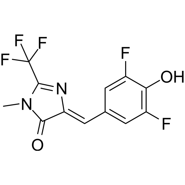 0 ImagesDFHBI-2T is a membrane-permeable, RNA aptamer-activated fluorescent probe (Ex/Em=500 nm/523 nm), with a Kd value of 1300 nM for the RNA aptamer Spinach2. DFHBI-2T binds to Spinach2, converting the fluorophore to a fluorescent state, stabilizing its planar structure to facilitate the radiative fluorescence decay pathway, and forming a complex with red-shifted excitation and emission peaks. DFHBI-2T can be used for RNA imaging in live cells. It is also applicable to research related to fragile X-associated tremor/ataxia syndrome.



